Art-labeling activity: structure of nervous tissue, a captivating technique, takes the stage as it illuminates the intricate architecture of the nervous system. This engaging exploration delves into the diverse types of nervous tissue, unveils the intricate structure of neurons, and unravels the crucial role of glial cells.
Join us on a journey to unravel the mysteries of the human nervous system through the lens of art-labeling activity.
Histological Structure of Nervous Tissue

Nervous tissue is a complex and specialized type of tissue that is responsible for transmitting information throughout the body. It is composed of two main types of cells: neurons and glial cells.
Neurons
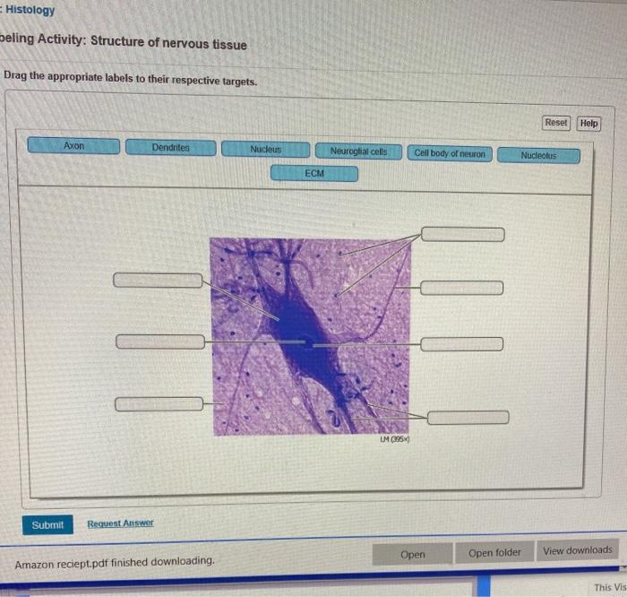
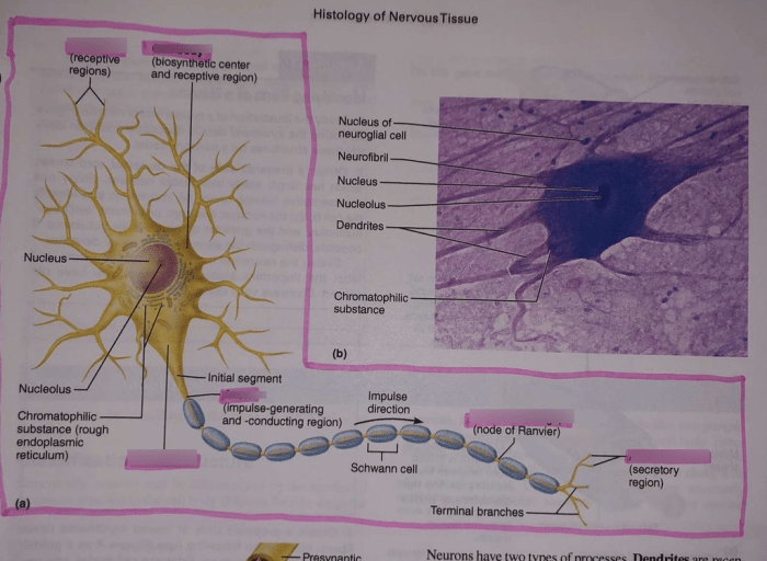
Neurons are the primary functional units of the nervous system. They are responsible for transmitting electrical and chemical signals throughout the body. Neurons have a cell body, dendrites, and an axon.
- The cell body contains the nucleus and other organelles that are necessary for the neuron’s survival.
- Dendrites are short, branched extensions of the cell body that receive signals from other neurons.
- The axon is a long, slender extension of the cell body that transmits signals to other neurons.
Glial Cells
Glial cells are non-neuronal cells that provide support and protection for neurons. They make up about 90% of the cells in the nervous system.
- Astrocytes are the most common type of glial cell. They help to maintain the blood-brain barrier and provide nutrients to neurons.
- Oligodendrocytes are responsible for myelinating axons, which helps to speed up the transmission of electrical signals.
- Microglia are the immune cells of the nervous system. They help to remove damaged cells and debris.
Methods for Labeling Nervous Tissue

There are a variety of methods that can be used to label nervous tissue. These methods allow researchers to visualize and study the structure of the nervous system.
Immunohistochemistry
Immunohistochemistry is a technique that uses antibodies to label specific proteins in nervous tissue. This technique can be used to visualize the distribution of specific proteins in the nervous system, such as neurotransmitters or receptors.
Histological Stains
Histological stains are dyes that can be used to label different types of cells and tissues. These stains can be used to visualize the structure of the nervous system, such as the location of neurons and glial cells.
Advantages and Disadvantages of Different Labeling Methods
Each labeling method has its own advantages and disadvantages. Immunohistochemistry is a highly specific method, but it can be time-consuming and expensive. Histological stains are less specific, but they are quick and easy to use.
Applications of Art-Labeling Activity

Art-labeling activity is a technique that can be used to study the structure of nervous tissue. This technique involves injecting a fluorescent dye into the nervous system and then using a microscope to visualize the labeled tissue.
Types of Art-Labeling Studies
There are a variety of different types of art-labeling studies that can be conducted. These studies can be used to visualize the structure of the nervous system, such as the location of neurons and glial cells, or to study the function of the nervous system, such as the flow of information through the brain.
Benefits and Limitations of Art-Labeling Activity
Art-labeling activity is a powerful tool for studying the structure and function of the nervous system. However, this technique also has some limitations. One limitation is that art-labeling activity can only be used to visualize living tissue. Another limitation is that art-labeling activity can be toxic to the nervous system.
Future Directions for Art-Labeling Activity: Art-labeling Activity: Structure Of Nervous Tissue

Art-labeling activity is a rapidly growing field of research. There are a number of potential applications for art-labeling activity in future research. One potential application is to use art-labeling activity to study the development of the nervous system. Another potential application is to use art-labeling activity to study the effects of drugs and toxins on the nervous system.
Challenges in Art-Labeling Activity, Art-labeling activity: structure of nervous tissue
There are a number of challenges that need to be addressed in order to advance the field of art-labeling activity. One challenge is to develop new art-labeling techniques that are less toxic to the nervous system. Another challenge is to develop new art-labeling techniques that can be used to visualize the structure and function of the nervous system in living animals.
Question & Answer Hub
What is art-labeling activity?
Art-labeling activity is a histological technique that utilizes specific antibodies or dyes to label and visualize different components of nervous tissue.
How does art-labeling activity aid in studying the nervous system?
Art-labeling activity allows researchers to identify and localize specific cell types, proteins, or structures within the nervous tissue, providing valuable insights into its organization and function.
What are the limitations of art-labeling activity?
While art-labeling activity is a powerful tool, it has limitations such as potential cross-reactivity of antibodies, tissue damage during processing, and the inability to label all components of the nervous system.